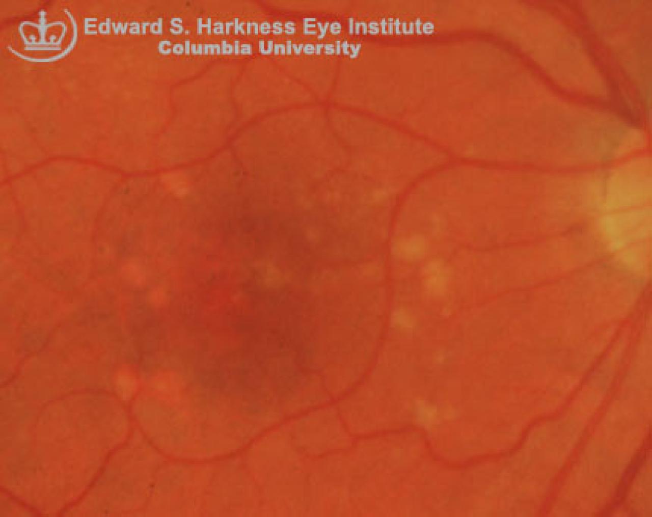
Have been referred to as "incipient atrophy" since they are usually preceding geographic atrophy and are more likely to develop soft drusen and choroidal neovascularization
Clinical Features
Areas of stippled, punctate hypopigmentation of degenerated RPE associated with pigment mottling
Thinning of the overlying neurosensory retina
The underlying choroidal vessels are not more readily apparent than in normal area without atrophy
Fluorescein angiogram demonstrates diffuse hyperfluorescence with a pattern of reticular or punctate blockage in the areas of RPE degeneration corresponding to the pigment clumping.