Pigment Dispersion Syndrome
- Mild pigment dispersion can be observed as a normal aging process
- Pigmentary glaucoma results when deposition of excessive pigment in the trabecular meshwork (as a result of liberation of pigment from the posterior iris epithelial surface in response to rubbing against the lens zonules) causes elevated intraocular pressure and subsequent optic disc damage
Clinical Features
- Symptoms may be asymptomatic or present as an intermittent or rapid onset of elevated intraocular pressure associated with corneal edema, ocular pain, intermittent blurring of vision or halos
- Signs:
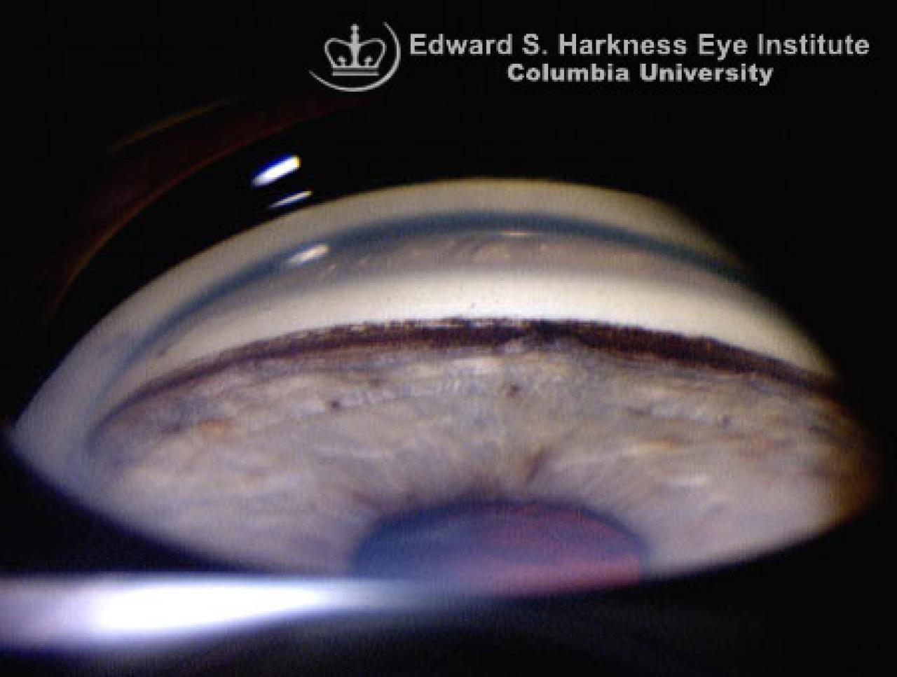
- Radial periphery transillumination defects of the iris
- Krukenberg spindle: a vertically oriented deposition of pigment on the posterior endothelial surface of cornea
- A band of dark brown or black pigment along the trabecular meshwork (on gonioscopy)
- Collection of pigment particles on the surface of the iris
- Dense pigment deposition on the posterior lens surface
- Posterior or concave bowing of the perpheral iris (on gonioscopy)
- Differential diagnosis may include the following: exfoliation syndrome, pseudophakia with malpositioned posterior chamber intraocular lens, iris or ciliary body cysts, intraocular pigmentary masses (e.g. melanoma), and uveitis
Management
- Observation and close follow-up of patients at risk
- Medical therapy if glaucoma present (or at risk for glaucoma)
- Surgical therapy includes:
- Peripheral laser iridotomy to flatten the iris and eliminate posterior bowing of the iris (thereby minimizing pigment liberation)