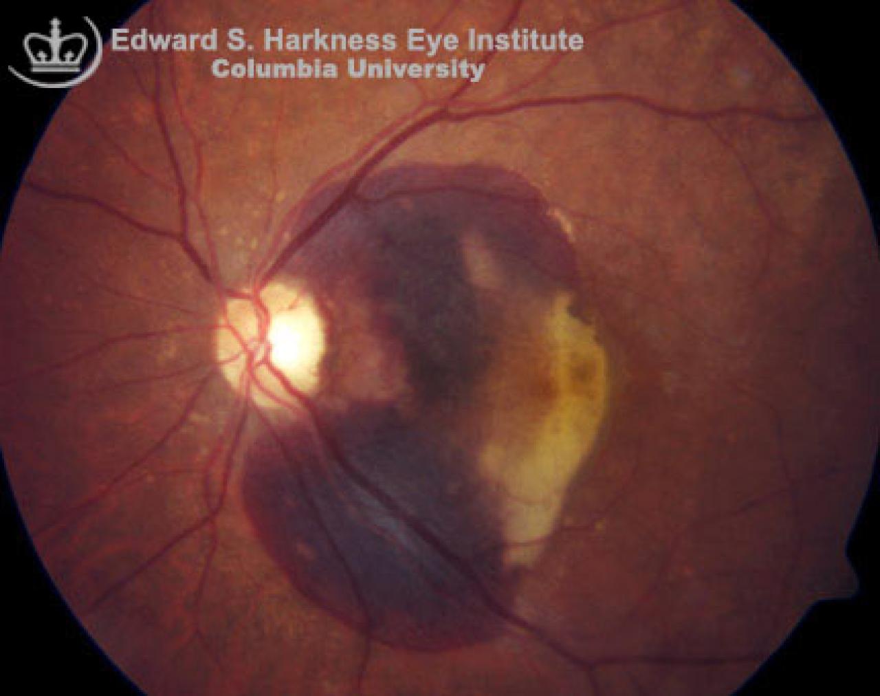
Situated deep to the retinal vessels and located under the retinal neurosensory level or RPE
Fluorescein angiography demonstrates:
Area of hypofluorescence, which is consistent with blockage of underlying details in areas of hemorrhages
Normal fluorescence of surrounding choroidal filling and overlying retinal vascular perfusion
Associated conditions include: choroidal neovascular membranes (CNVM), trauma that lead to choroidal rupture, macroaneurysms or exudative age-related macular degeneration.