Splinter Optic Disc Hemorrhage
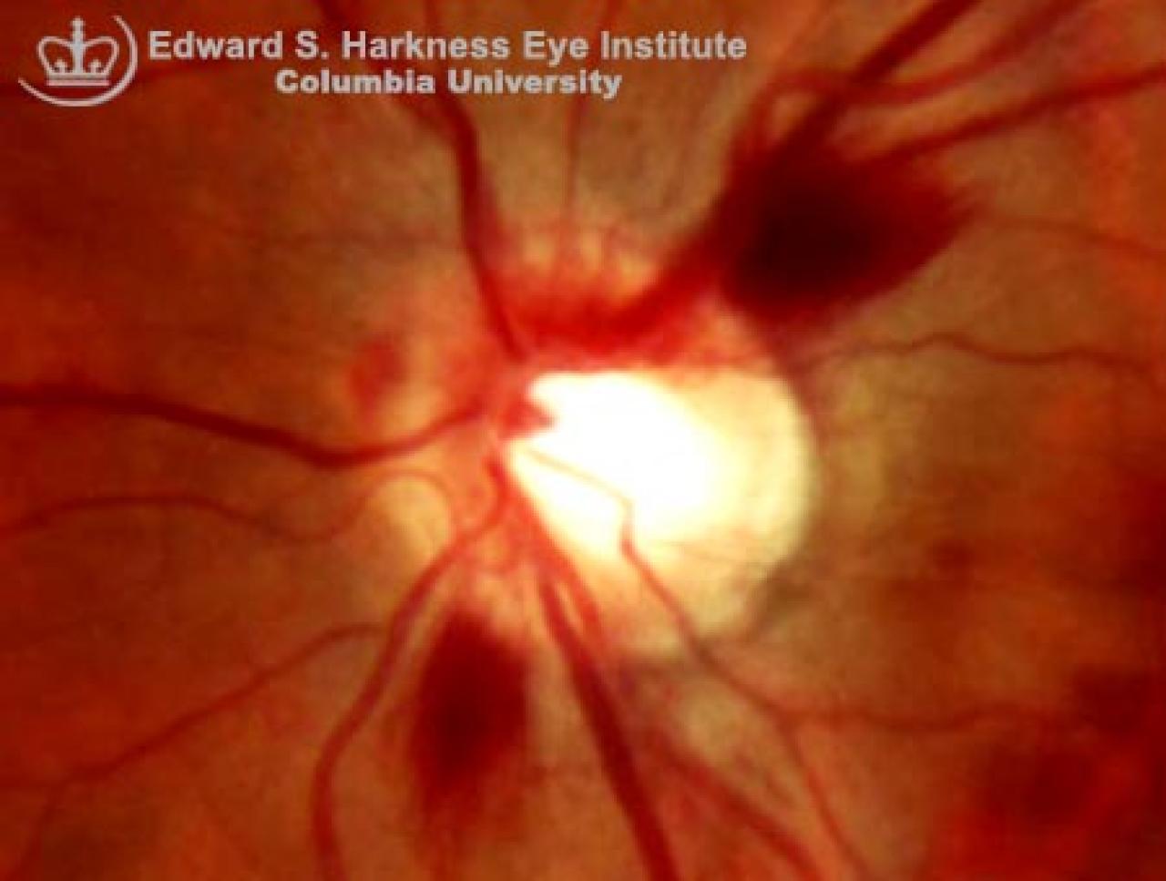
Glaucomatous optic disc demonstrating:
- Increased cupping and central pallor with baring of circumlinear vessel
- Splinter optic disc hemorrhages
- Nasalization of the vessels
- Localized notching of the neural rim between the 3 and 4 o'clock position
- Diffuse thinning of the retinal nerve fiber layer is evident with increased visibility of small vessels and capillaries