Degenerative Myopia
- Progressive retinal and choroidal degeneration in myopic eyes of at least - 6.00 Diopter refractive errors with an axial length of more than 26 mm.
- Progressive chorioretinal stretching results in characteristic fundus findings :
- Optic disc crescent, area of depigmentation due to dragging of the choroids and RPE from the optic nerve
- Ruptures of Bruch's membrane (lacquer crack)
- Focal areas of chorioretinal atrophy
- Localized ectasia involving the sclera, the pigment epithelium and the choroid (posterior staphyloma)
- Oval with vertical axis and slightly tilted optic disc
- Localized areas of pigment epithelial proliferation (Foerster-Fuchs spots)
- Lattice degeneration in the pre-equatorial area may be found
- Thinning of the RPE and the choriocapillaris give a 'tigroid' or 'tesselated' fundus appearance
Complications
- Earlier onset posterior vitreous detachment
- Small macular hemorrhage
- Macular hole
- Posterior retinal detachment
- Choroidal neovascularization (CNV)
- Fluorescein angiogram is helpful to reveal choroidal neovascularization.
Management
- Laser photocoagulation for selected cases of CNV
- Posterior scleral buckling for posterior retinal detachment

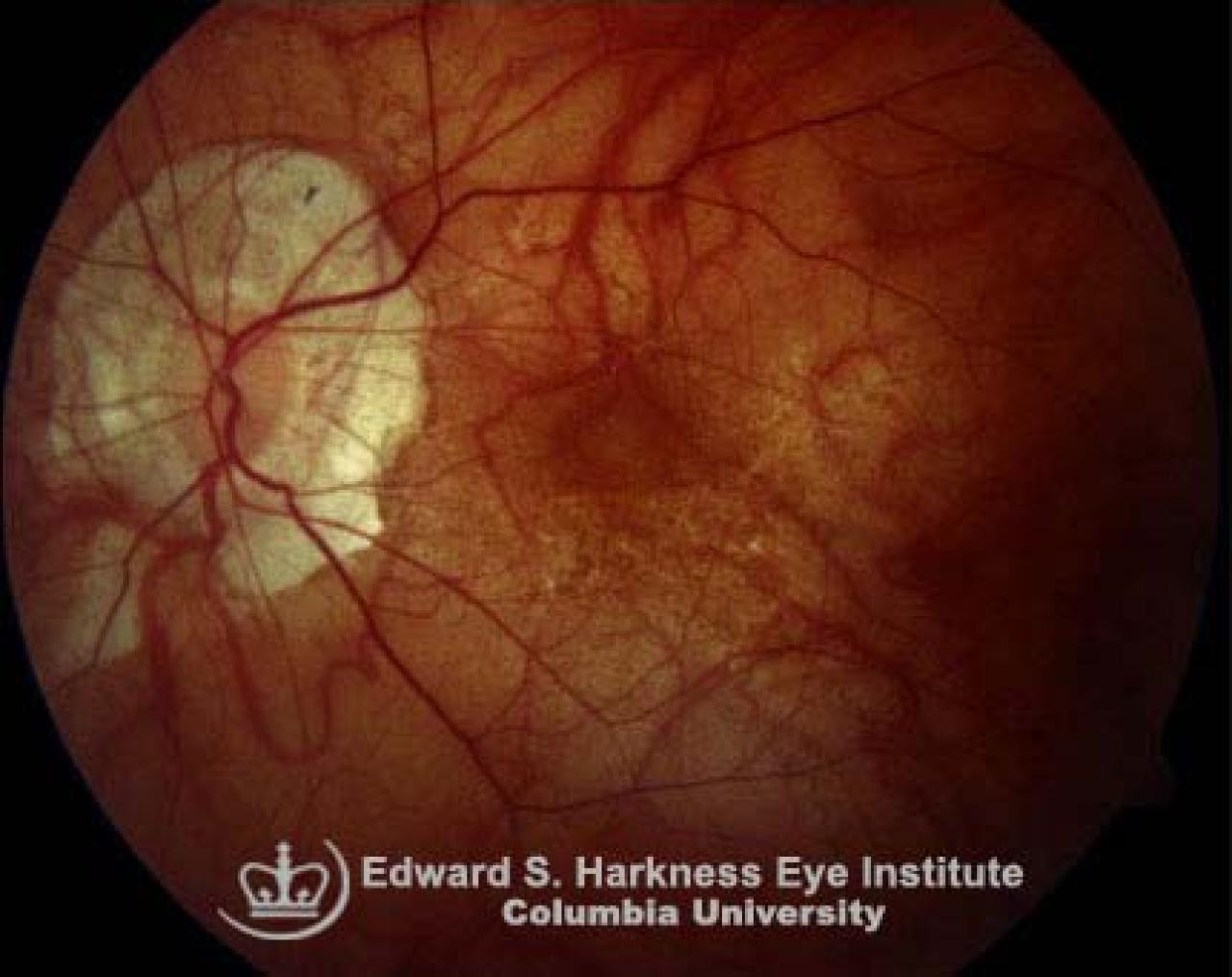
Typical features of degenerative myopia demonstrating:
- Peripapillary atrophy
- Tilted disc
- Tesselated or tigroid fundus
- Multiple areas of pigment epithelium and choriocapillaris atrophy involving the macular area
- Lacquer cracks
- Pigment epithelium proliferation produces Foerster-Fuchs' spots