Symptoms: usually asymptomatic, but may affect vision over time due to progression of streaks towards the fovea
Signs:
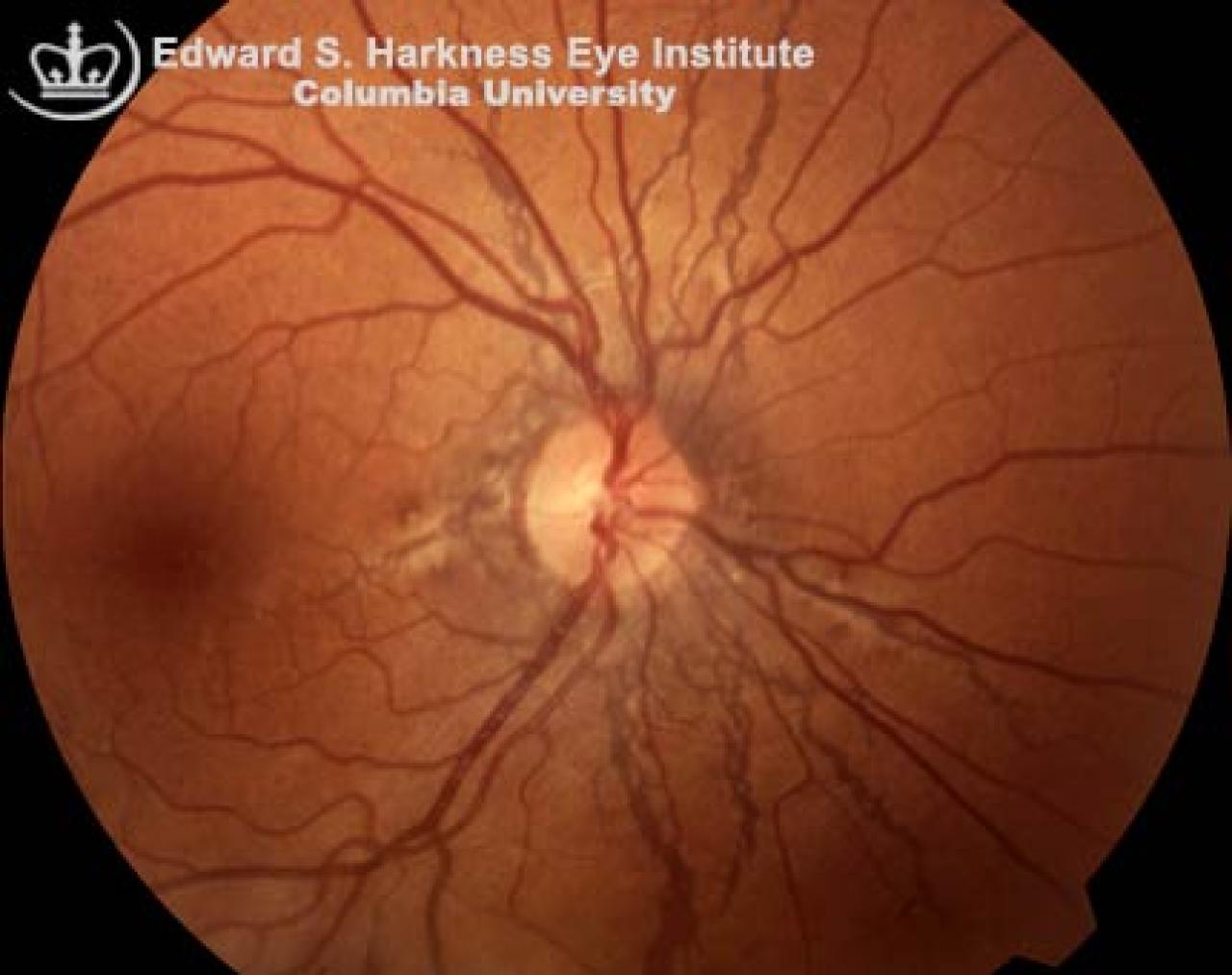
Irregular, spokelike, curvilinear or jagged streaks that radiate outward from the peripapillary area towards the peripheral fundus or can be concentric to the optic disc
Near the optic disc, they may be interconnected by circular breaks
Color varies from reddish orange to dark brown, or appear grayish if fibrovascular tissue is present
Associated funduscopic findings may include:
Peau d'orange (orange skin) pattern of diffuse mottling of the pigment epithelium in the temporal midperiphery
Peripheral subretinal crystalline bodies
Focal atrophic spots
Optic nerve drusen
Fluorescein angiographic findings:
Irregular hyperfluorescence of the streaks during early phases and late staining
Can be seen as hypofluorescence of the streaks outlined by hyperfluorescence margins, which stain in the late phases
Some clinically invisible streaks may be observed during fluorescein angiography
Most common associated systemic conditions:
Idiopathic
Pseudoxanthoma elasticum (PXE)
Paget's disease
Sickle cell disease
Ehler's- Danlos Syndrome
Complications
Choroidal neovascularization
High risk of severe subretinal hemorrhages due to rupture of the Bruch's membrane following a relatively mild ocular injury
Management: laser photocoagulation in selected cases of choroidal neovascularization, but the recurrence rate is high.