Central Retinal Artery Occlusion (CRAO)
- Typically unilateral.
- More commonly affect older people in their mid-sixties, but can also occur in younger patients.
- Most common cause: systemic hypertension.
- Other etiologies: diabetes mellitus, emboli from valvular heart diseases, carotid atherosclerosis and DVT, circulatory compromise, coagulopathies, collagen vascular diseases, other vasculitides and trauma.
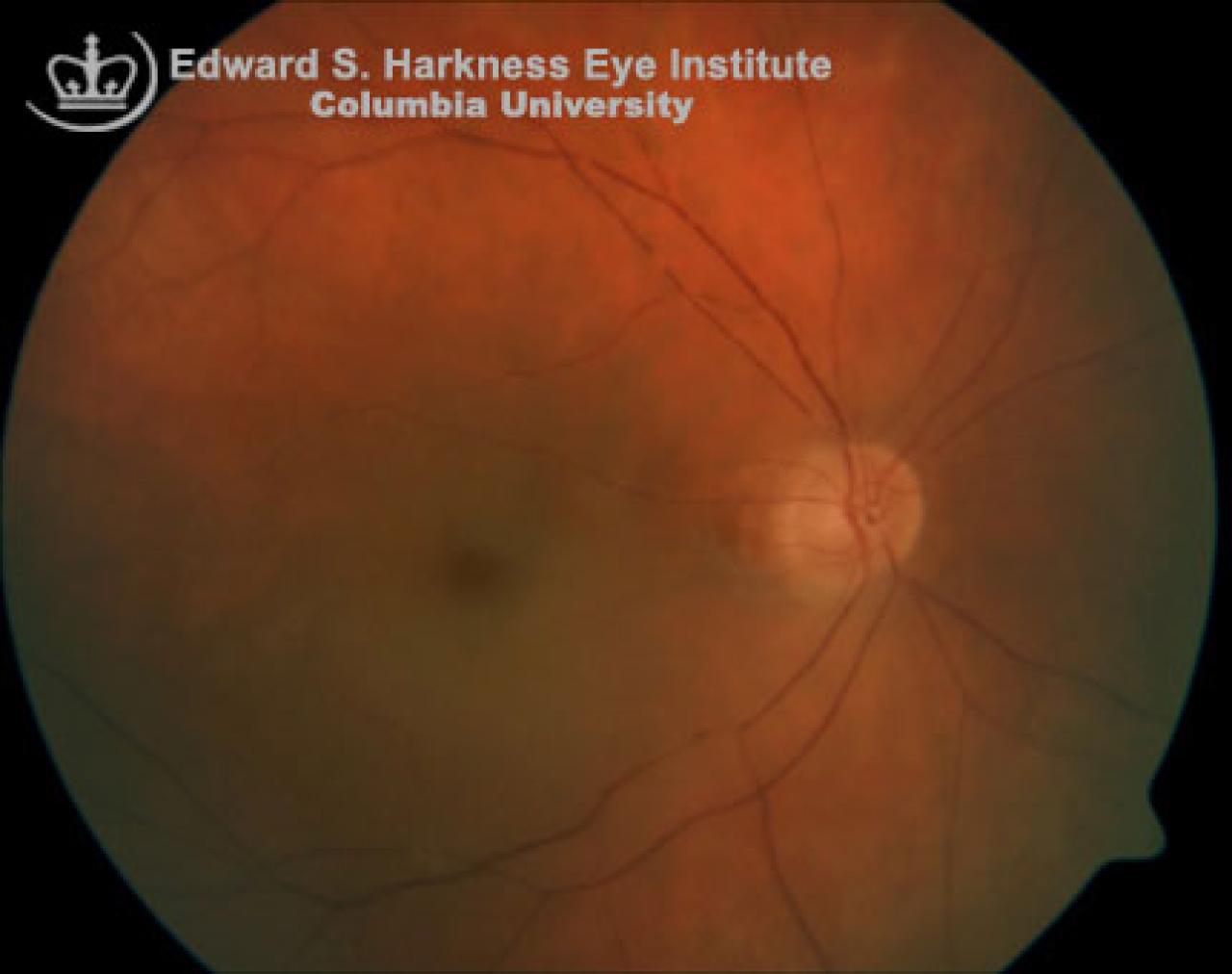
Clinical Features
- Symptoms:
- Sudden, painless loss of vision
- May have a history of amaurosis fugax
- Signs:
- Normal anterior segment in acute cases
- Pale, whitening, swelling retina especially in the posterior pole
- Cherry red spot as a presentation of orange reflex from the intact choroidal vasculature beneath the foveola surrounded by the retinal pallor
- Afferent pupil defect is usually present
- Emboli may be seen
- After 4-6 weeks, the cloudy swelling retinal commonly resolves, leaving a pale optic disc, attenuated retinal vessels, segmentation or "boxcarring" of the blood column
- In most cases, neovascularization of the iris usually present by this time
- Final visual acuity is most often worse than 20/400
- Visual acuity of better than 20/40 may be achieved with patent cilioretinal artery
Fluorescein Angiography Demonstrates
- Delay in retinal arterial filling and arteriovenous transit time
- Segmentation of the blood column
- Choroidal vascular filling is usually normal
Management
- Thorough evaluation of systemic etiology.
- May consider the following treatment to lower the intraocular pressure: ocular massage, anterior chamber paracenthesis.
- Other treatments may include: oral vasodilator and systemic anticoagulants.
- Panretinal photocoagulation in the presence of iris neovascularization.